
Anxiety is a common negative emotion and it is also one of the buzzwords in today’s society. When we suffer from anxiety, what changes will occur in our brains? The research team led by Prof. LI Xiaoming at the Zhejiang University School of Medicine conducted an in-depth study regarding this. They found that different anxiety-related behaviors are mediated by different neural circuits and molecular mechanisms.
Their research findings were published in a cover article entitled “Distinct serotonergic pathways to the amygdala underlie separable behavioral features of anxiety” in the journal Nature Neuroscience on November 29.
The amygdala, located in the medial temporal lobe, is the brain region primarily associated with emotional processes. The name amygdala is derived from the Greek word amygdale, meaning “almond,” owing to the structure’s almond-like shape. Previous studies have revealed that the amygdala plays a crucial role in controlling fear, reward and depression in mice. As a part of the “emotional brain”, the amygdala is evolutionally conservative. Thus, studies from the mouse brain will help us gain a better understanding of the human brain. To this end, LI Xiaoming’s team has been long committed to researching into the neural circuits of the amygdala and the neural mechanisms for emotions such as fear, anxiety and depression (Nature Medicine 2019, Nature Neuroscience 2019, Neuron 2019, 2020, Cell Research 2016, eLife 2018, 2020, Neuroscience Bulletin 2020, 2022, Journal of Neuroscience 2017).
“Anxiety is an emotional response to vague and potential threat, as manifested by a series of physiological reactions and avoidance,” said YU Xiaodan, the first author of the study. Chronic exposure to anxiety will deteriorate into anxiety disorders, represented by extremely anxious state and accompanied social deficits. Mice exposed to anxiogenic stimuli spend more time in ‘safe’ zones of the behavioral apparatus and spend less time interacting with an unfamiliar conspecific. However, how does the brain integrate anxiogenic information and mediate anxiety-related behaviors is still unclear.
In mice, the midbrain dorsal raphe nucleus (DRN) contains the majority of forebrain-projecting 5-HT neurons, which regulate emotional and motivational processes. DRN5-HT neurons innervate the basolateral amygdala, especially the basal nucleus, also known as the basal amygdala (BA). Previous studies demonstrate that the BA regulates the development of fear to some extent, but does it induce anxiety and the different behavioral features of anxiety?
To answer these questions, the research team used their self-developed genetically encoded GPCR-based sensor for 5-HT (GRAB5-HT2h) to record the dynamics of extracellular 5-HT levels in the BA in social or anxiogenic conditions. 5-HT in the BA increased in social condition, but decreased in the anxiogenic state. However, in both situations, DRN5-HT neurons were activated. Given that the activation of DRN5-HT neurons would prompt the release of 5-HT and that the level of 5-HT in the BA displayed opposite dynamics in anxiety and social conditions, this seemingly paradoxical phenomenon inspired the researchers to investigate its neural mechanisms. They speculated that there would be heterogeneity between DRN5-HT neurons and the BA, thereby resulting in the decrease of 5-HT in the BA in the anxiogenic state.
In their study, the researchers found two subpopulations of 5-HT neurons: DRNvGluT3∩5-HT-BAPV and DRNvGluT3∩5-HT-BAPyr neurons, which possess distinct features in electrophysiological intrinsic properties, gene expression and responses to anxiogenic and social stimuli. Specifically, DRNvGluT3∩5-HT-BAPyr neurons and DRNvGluT3∩5-HT-BAPV neurons were activated by social and anxiogenic stimuli, respectively. Activation of the DRNvGluT3∩5-HT→BAPV pathway excited BAPV neurons via glutamate. The robust GABA efflux from BAPV neurons inhibited 5-HT release across the BA via GABAB receptors. The reduction of 5-HT in BA induced avoidance of bright spaces and social avoidance, with avoidance of bright spaces mediated by HTR1A and social avoidance being mediated by HTR1B. These results reveal the DRNvGluT3∩5-HT-dependent precise control of BA neurons in the regulation of different behavioral features of anxiety.
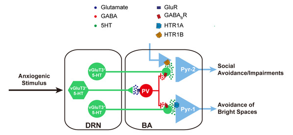
DRNvGluT3∩5-HT→BAPyr and DRNvGluT3∩5-HT→BAPV pathways differentially regulate anxiety-related behaviors.
This is the first time that scientists have discovered the elaborate neural circuit mechanisms and the specific neuromolecular basis of different anxiety-related behaviors, from the perspective of behavioral phenotypes. This research provides novel approaches and a theoretical foundation concerning the occurrence and pathogenesis of anxiety disorders through the symptomatology. In addition, this study reveals the heterogeneity of 5-HT neurons and amygdala glutamatergic neurons from a new perspective—the heterogeneous projection and cell-type specific connectivity, further explains the functional diversity of 5-HT, and expands the current understanding of the structure and function of 5-HT and the amygdala.
Image credit: The research team led by Prof. LI Xiaoming; Nature Neuroscience
(From ZJU NEWSROOM)