Conventional experiments in chemistry and biology study the behavior of the ensemble, but it has been an abiding scientific challenge for scientists to observe, manipulate and measure the chemical reactions of individual molecules.
In response to this challenge, Prof. FENG Jiandong from the Department of Chemistry of Zhejiang University has committed himself to developing interdisciplinary single-molecule techniques and instruments to observe single-molecule chemical reactions in solution. Recently, Feng and his colleagues have devised a novel technique for directly imaging single-molecule electrochemical reactions in solution with ultrahigh spatial resolution. This technique shows important applications in the fields of chemical imaging and biological imaging such as imaging microstructures and cells with nanometer resolution. The research finding is published as a cover story of the August 11 issue of Nature.

In comparison with fluorescence imaging, electrochemiluminescence (ECL) imaging does not require the use of excitation light, so there is minimal background. ECL is an important tool in in vitro immunodiagnosis which requires ultrahigh sensitivity for resolving weak signals. At present, there are two major challenges in the ECL field. First, it is vitally important for single-molecule assays that ECL signals can be measured and imaged at weak or even single-molecule level. Second, it is of tremendous significance to chemical and biological imaging if super-resolution ECL microscopy—ultrahigh spatiotemporal imaging which breaks the optical diffraction limit—can be developed.
Over the past three years, Feng and his team have been working on these two major problems. They developed a combined widefield optical imaging and electrochemical recording system and built an efficient ECL control, measurement and imaging setup. They performed the first widefield imaging of single-molecule ECL reactions and on the strength of this, they achieved the first super-resolution ECL imaging. Without any light excitation, this single-molecule ECL microscopy can achieve single-molecule super-resolution imaging, which has great potential for applications in chemical measurements and biological imaging.
Why is it hard to spatially capture single-molecule signals during the ECL process? It is primarily attributed to the fact that single-molecule reactions are difficult to control, track and detect. “Single-molecule chemical reactions are accompanied with exceedingly weak optical, electrical and magnetic signal changes, and the process of chemical reactions and the location where chemical reaction occurs are stochastic,” said Feng.
To this end, Feng and his colleagues built a sensitive detection system which can capture luminescence signals generated after single-molecule reactions. “Imaging single reactions calls for the spatial and temporal isolation of individual reaction events,” said Feng. “This is achieved in our case by using diluted solutions and fast camera acquisitions,” said DONG Jinrun, a PhD candidate of the research team.
Microscopy is a crucial tool in material science and life science. Conventional optical microscopy works on the scale of hundreds of nanometers and beyond while high-resolution electron microscopy and scanning probe microscopy can reveal objects down to the atomic scale. “At this scale, there are still very limited number of technologies available for in situ, dynamic and solution observations at length scales ranging from a few nanometers to hundreds of nanometers,” said Feng, “This has a lot to do with inadequate optical imaging resolution due to the optical diffraction limit.” Accordingly, the team started to work on super-resolution ECL imaging by spatiotemporally isolating single-molecule signals.

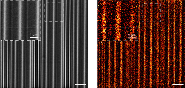
(left) Scanning electron microscopy images for focused ion beam (FIB) patterned ITO
(right) Super-resolved single-molecule ECL images for the same FIB patterned ITO
Inspired by super-resolution fluorescence microscopy, they employed the optical reconstruction of localized spatial molecular reactions for imaging. This is similar to how one can distinguish between two adjacent stars at night by their “twinkling” behavior. “The spatial localization of luminescence sites and the super-imposition of information regarding each frame of isolated molecular reaction sites constitute a ‘constellation’ of chemical reaction sites.”
To attest to the feasibility of this imaging method and the accuracy of the localization algorithm, the team fabricated a pattern of stripped electrode as a known imaging template and conducted comparative imaging. The results of single-molecule ECL imaging agreed well with the results of electron microscopy imaging in structure, verifying the feasibility of this imaging method. Single-molecule ECL imaging increased the spatial resolution of conventional ECL microscopy to an unprecedented 24 nanometers.
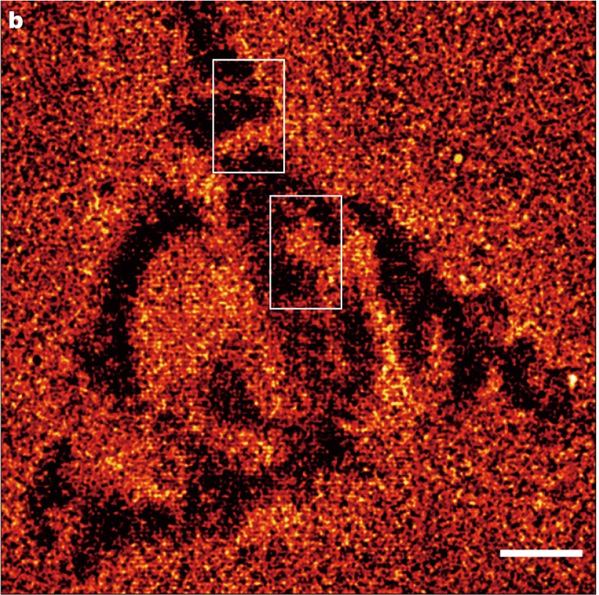
FENG Jiandong and his colleagues went on to apply single-molecule ECL imaging to cell imaging. There was no need for direct labeling for ECL cell imaging, which may be potentially friendly to cells, as the traditional labeling process may affect the cell state. They further performed single-molecule ECL imaging on cell adhesions and observed their dynamics over time. By comparing the correlated ECL imaging and super-resolution fluorescence imaging results, they found that ECL imaging exhibited high spatial resolution comparable to super-resolution fluorescence microscopy while avoiding the use of lasers and cell labeling.

Super-resolved ECL image of a single live cell
“The authors’ findings open the way to a new concept in imaging: a chemistry-based approach to super-resolution microscopy,” Prof. Frédéric Kanoufi from the University of Paris and Prof. Neso Sojic from the University of Bordeaux wrote in an accompanying commentary in Nature journal's news and views. “It could also lead to the development of new strategies for bioassays and cell imaging, complementing well-established fluorescence-based single-molecule microscopy techniques.”
Image credit: Nature
(From: ZJU NEWSROOM)

